Which Disease is it? Sunday, Jan 1 2017
Diagnostics has been a central part of clinical medicine right from the beginning. It’s not hard to see why—how can a patient be treated properly without a clear idea of what the condition is? Diagnosing the problem is so fundamental that even the cranks and the nutters must also pay some sort of deference to it. It follows that the more precisely the condition can be diagnosed, the better crafted and thus more personalised, the treatment can be. Our understanding of the workings of human body has been established at a molecular level, as has our understanding of some disease.
The simplest forms of these are well known: the presence of a given metabolite in the blood or urine are obvious examples. Conditions such as diabetes and even pregnancy can be identified readily. These two are relatively straight forward to diagnose using metabolites: the presence of really any concentration of glucose or human chorionic gonadotrophin (hCG) is sufficient to indicate a positive identification.
However, not all conditions are similarly black-or-white. Some systems depend upon a development of tissues in order to function properly, for example in lungs. In order for a baby to be born and breathe properly thereafter, its lungs must of course develop sufficiently. However, assessing this is a challenge; how mature does a lung need to be in order to function? This is an important question as of course there is little time to do anything about poor lung function once the baby has been born.
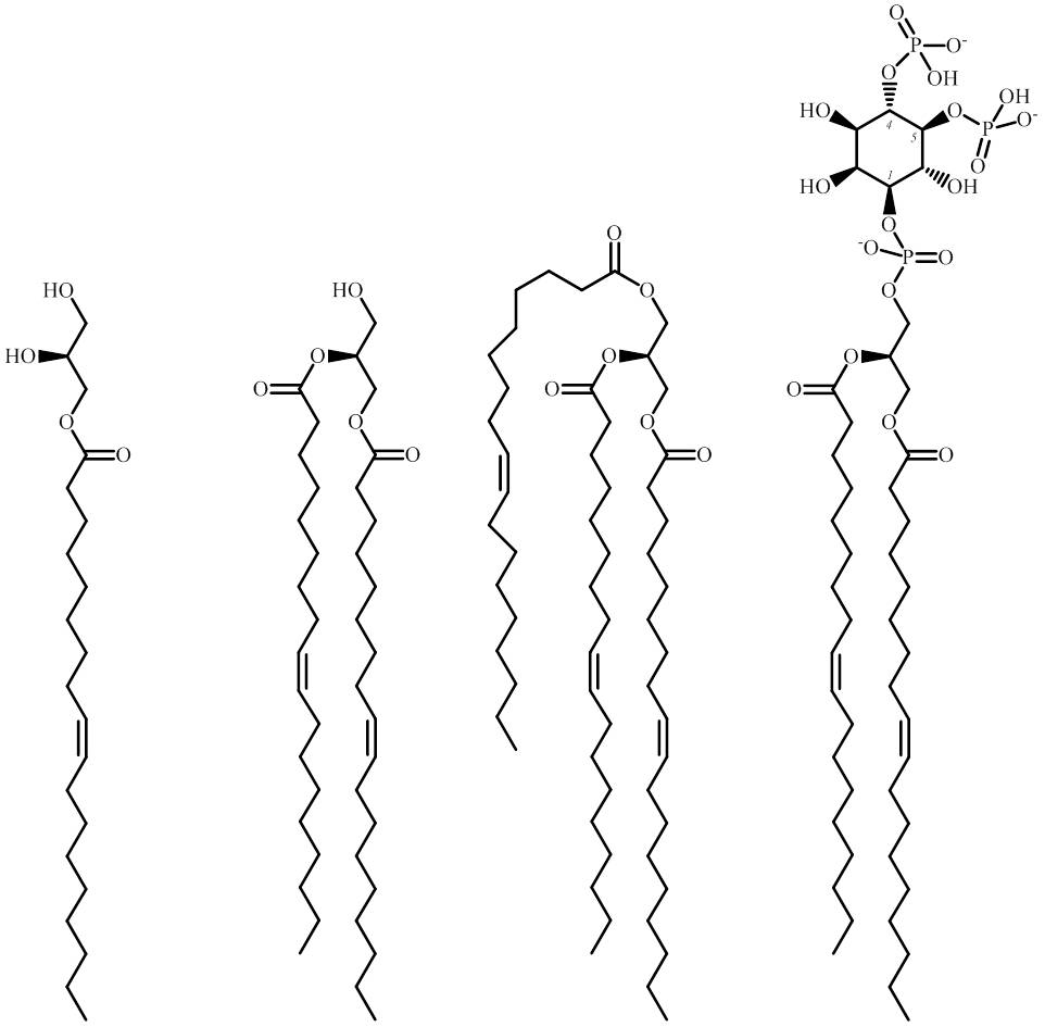
A recent collaboration between several laboratories in Scandinavia and the UK has produced a way of testing for lung maturity before it is too late, by profiling the lipid composition of gastric aspirates [1]. This has allowed prediction of Respiratory Distress Syndrome with a high degree of accuracy, that may yet be used clinically.
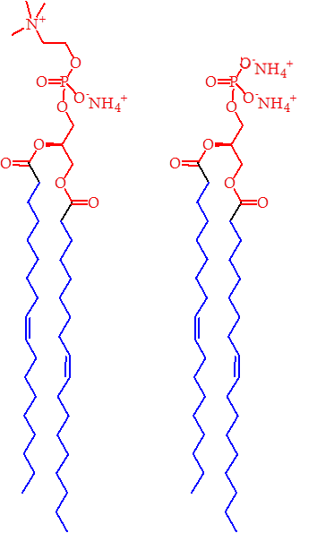
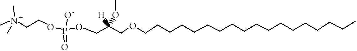
A quick medical response is also required with many head injuries. Such injuries are well-known in contact sports such as ice hockey, but conditions such as concussion can be difficult to diagnose in the early stages, despite long-term neurological consequences for the patient. A recent study in Canada showed that the abundance of isoforms of PC are modulated in at least 90% of cases of concussion [2]. This offers an opportunity for both an additional diagnostic test and also a means for understanding how cell structure may change as a result of such an injury.
A third recent development may also assist in diagnosing injury, though it is one that can vary enormously between individuals. Lipid profiling has been used to predict clinical outcomes in patients suffering from burns [3]. It appears that increases in the abundance of unsaturated fatty acids in blood plasma correlate with death of the patient. It is not yet clear what preventative measures can be taken with these data, however, it seems that like the lipid profiling of gastric aspiration, lipid profiling of burns victims may give an insight that a visual examination may not and thus inform medics about the need for preventative treatment.
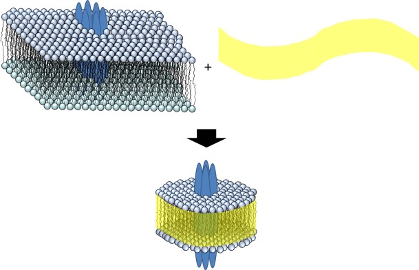
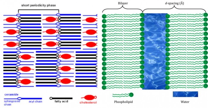
That lipid profiling may be used as a diagnostic tool in three quite separate conditions hints that it may be possible to use it in a host of other conditions, if only we know which lipids or lipid ratios to look at. These three studies have all required a healthy dose of serendipity and scientific intuition—as well as hard work—to find out what they have. We may not need to rely upon this nous to find all such examples, however. Identification of shifts in the lipid fraction that provide diagnostic evidence for other conditions may be informed by the availability of novel techniques that allow us to understand which lipids are where [4] and deeper analyses of lipid distribution [5].
References
[1] H. Verder, C. Heiring, H. Clark, D. Sweet, T. E. Jessen, F. Ebbesen, L. J. Björklund, B. Andreasson, L. Bender, A. Bertelsen, M. Dahl, C. Eschen, J. Fenger-Grøn, S. F. Hoffmann, A. Höskuldsson, M. Bruusgaard-Mouritsen, F. Lundberg, A. D. Postle, P. Schousboe, P. Schmidt, H. Stanchev, L. Sørensen. Acta Pædiatrica, 2016, DOI: 10.1111/apa.13683.
[2] M. Daley, G. Dekaban, R. Bartha, A. Brown, T. Charyk Stewart, T. Doherty, L. Fischer, J. Holmes, R. S. Menon, C. A. Rupar, J. K. Shoemaker, D. D. Fraser. Metabolomics, 2016, DOI: 10.1007/s11306-016-1131-5.
[3] P. Qi, A. Abdullahi, M. Stanojcic, D. Patsouris, M. G. Jeschke. Scientific Reports, 2016, DOI: 10.1038/srep38707.
[4] G. Lia, J. H. Kima, Z. Huanga, J. R. St. Claira, D. A. Browna, E. London. Proceedings of the National Academy of Sciences, 2016, DOI: 10.1073/pnas.1610705113.
[5] C. L. Jackson, L. Walch, J. M. Verbavatz. Developmental Cell, 2016, DOI: 10.1016/j.devcel.2016.09.030.


